ISO 9001
CE Certified
Make In India
Features
- How It Works (Simplified)
- Common Uses
- Diagnosing bone fractures, dislocations, arthritis.
- Checking for lung infections (pneumonia) or chest issues.
- Detecting foreign objects.
- Dental evaluations (cavities, tooth issues).
- Imaging the digestive tract (with contrast).
Technical Specifications
| Radiography/Plain Film | The process of using X-rays to produce images, or the resulting image itself. |
| Radiograph | The actual picture (black-and-white image) of the body's interior. |
| Ionizing Radiation | The high-energy electromagnetic waves used, similar to light but with more energy. |
| Radiologist | A doctor who interprets the X-ray images. |
| Radiographer/Technologist | The healthcare professional who operates the machine and takes the images. |
| Attenuation | The process where body tissues absorb X-rays, with denser tissues absorbing more. |
| Contrast Media | Substances (like barium or iodine) sometimes given to highlight specific organs or vessels. |
| Generation | An X-ray tube generates high-energy photons (X-rays). |
| Exposure | The beam passes through the patient, who is positioned between the source and a detector. |
| Absorption | Bones block most X-rays (white), while air lets them pass (black), with soft tissues in between (shades of gray). |
| Image Capture | The X-rays reaching the detector create the radiograph, traditionally on film, now often digitally. |
Similar Products
View all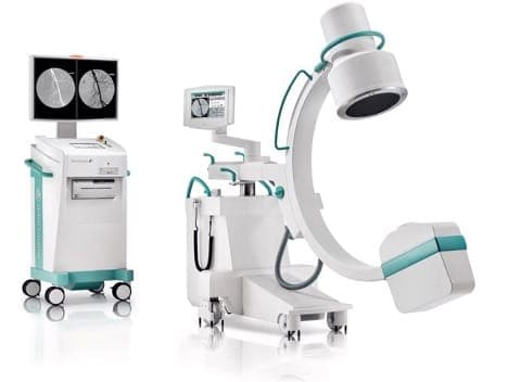
C-ARM MACHINE
Technology: Uses X-ray fluoroscopy to generate continuous, live images, not just static X-rays., Nam...
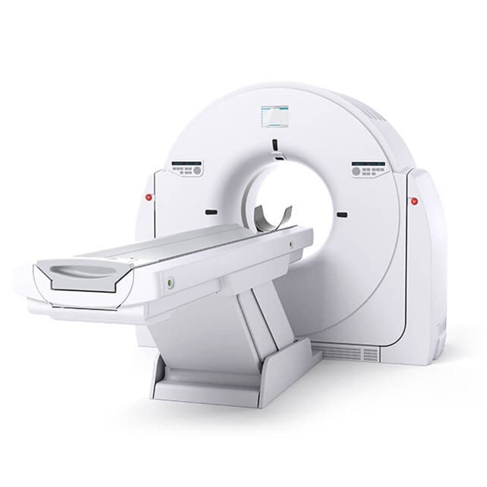
CT SCAN MACHINE
A Computed Tomography (CT) scan is a medical imaging procedure that uses specialized X-ray equipment...
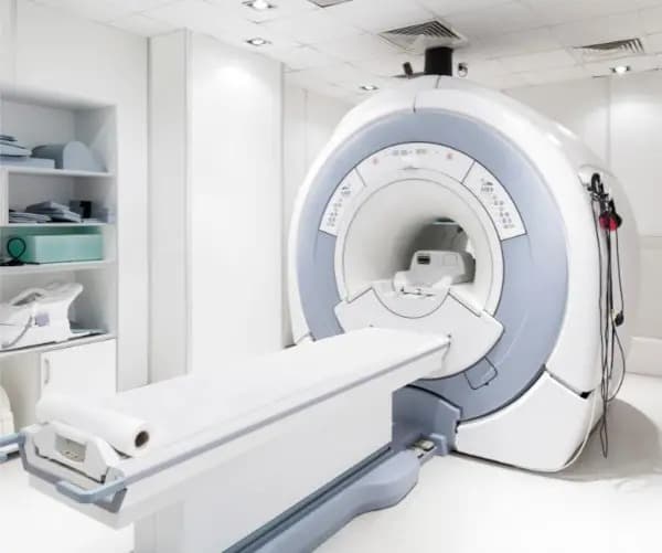
MRI MACHINE
A Magnetic Resonance Imaging (MRI) scan is a non-invasive medical imaging technique that uses strong...