ISO 9001
CE Certified
Make In India
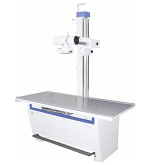
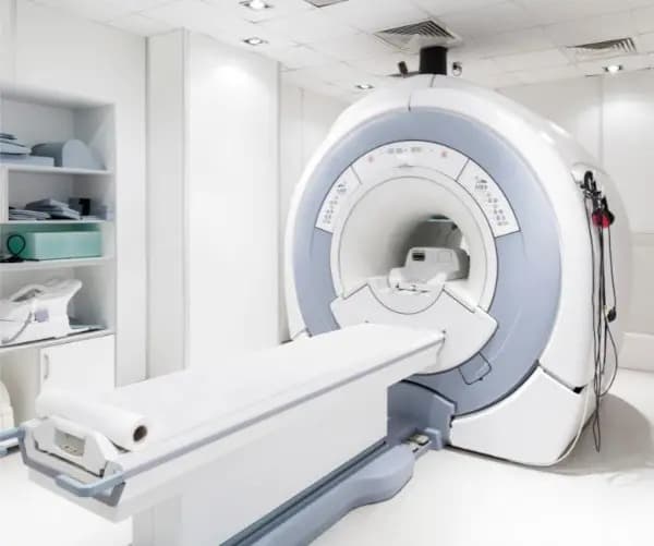
Technical Specifications
| Gantry | The donut-shaped machine housing the X-ray tube and detectors. Key specs include an aperture (opening) of at least 70-75 cm and a rotation time of less than 0.75 seconds (sometimes as fast as 0.35 seconds for high-end models). |
| X | ray Generator & Parameters: |
| Power Output | 32 KW or higher (up to 100 kW or more for high-end units). |
| Tube Voltage (kVp) | Range of 80-140 kV. |
| Tube Current (mA) | Range of 10 mA to 800 mA or more. |
| Detectors | Modern systems use solid-state detectors arranged in multiple rows to acquire multiple slices per rotation (e.g., 64, 128, or more physical rows). |
| Slice Thickness | Reconstructed slice thickness can range from sub-millimeter (as low as 0.4-0.63 mm) to 10 mm, allowing for high-resolution imaging. |
| Scanning Modes | Includes both axial (step-and-shoot) and helical/spiral scanning modes, with variable pitch factors (typically 0.3-1.5). |
| Reconstruction Matrix | Standard matrix sizes are 512x512, 768x768, and 1024x1024 pixels. |
| Reconstruction Speed | Capable of real-time reconstruction, often 20 images per second or more. |
| Hounsfield Units (HU) | Images display tissue density on the Hounsfield scale, where water is 0 HU, air is -1000 HU, and bone is typically +400 HU or more. |
| Patient Table | Motorized table made of carbon fiber with a high weight capacity (typically ≥ 200 kg) and accurate positioning. |
| Software | Advanced features often include multi-planar reconstruction (MPR), 3D volume rendering (VR), maximum intensity projection (MIP), metal artifact reduction (MAR), and low-dose protocols for pediatric patients |